Where Is the Sternum in Relation to the Shoulder?
THORACIC SPINE REHABILITATION
INTRODUCTION & anatoMY
Middle back pain or stiffness is a common problem. This is especially true in today's society where prolonged sitting and reduced physical activity are increasingly commonplace. In most cases people experience either acute pain or long-standing stiffness in their thoracic spine, and in some cases symptoms may fluctuate and be recurrent in nature. Problems in the middle back can also be related to the ribs and the chest (Sternum) that it attaches to, and the overlying shoulder blades. Having a good understanding of the anatomy of the middle back, the source of your symptoms and what they mean will help you to progress and make a good recovery.

The Middle Back (Thoracic Spine) refers to the middle 12 vertebrae where our ribs attach
Complete Anatomy Version 2.3.7 (4120), 2017 (1)
ANATOMY OF THE MIDDLE BACK (THORACIC SPINE)
The middle back - called the 'thoracic spine' - refers to the middle portion of our spine, beginning at the base of our neck and ending to the bottom of our rib cage. It comprises 12 thoracic vertebrae, spaced apart by intervertebral discs, and connected by facet joints and ligaments. The facet joints are oblique and flattened to allow more rotational movement in this part of the spine. Inside the vertebral arch lies the spinal cord, and nerve roots exit between each of the vertebrae. These nerve roots travel to provide sensation and muscle contraction in our upper limbs and torso and innervation to our internal organs. They also merge to form a collection of nerves known as the sympathetic and parasympathetic nerve trunks which help to maintain normal equilibrium in our body. In conjunction with brain and hormonal system function these nerve trunks control whether our body aims to rest and digest or to be prepared for physical activity and exertion.
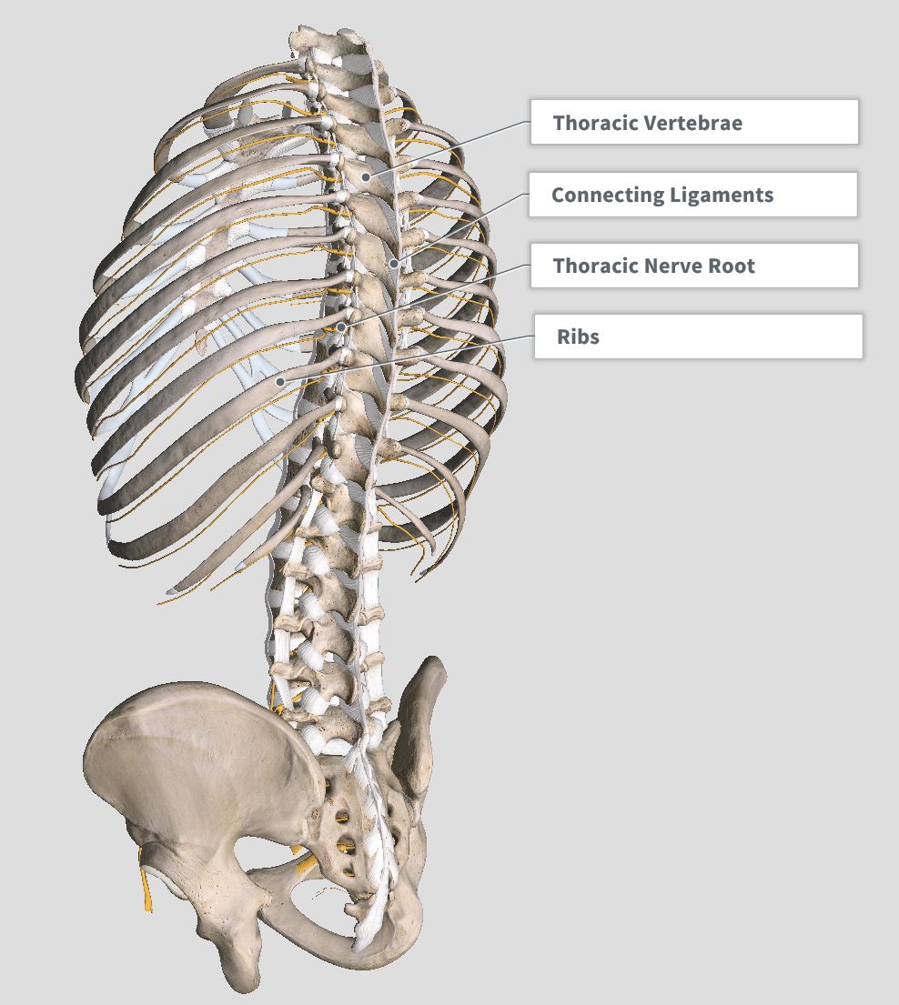

The spine is comprised of vertebrae, spaced apart by inter-vertebral discs, and connected by facet joints and ligaments. Spinal cord tissue is enclosed in the vertebral arch. Nerve roots exit in the spaces between vertebrae. The middle back - 'thoracic spine' refers to our middle 12 vertebrae surrounded by our rib cage. Complete Anatomy Version 2.3.7 (4120), 2017 (1)
Connected to the sides of the thoracic vertebrae are 12 pairs of ribs. The ribs extend around to connect to our chest plate (sternum), forming successive rings of bone in our thorax that protect the heart and lungs and function in breathing. The ribs have small joints and ligaments that connect them to the sides of the vertebrae (costotransverse joints) and to the bodies of the vertebrae (costovertebral joints). The ribs also have joints that connect them to our sternum (sternocostal joints). Large portions of the ribs near our chest wall are made of cartilage rather than bone, and where this transition from cartilage to bone occurs there is small connections called costochondral joints.


In the thoracic spine, the ribs connect up to the vertebrae and to the sternum via the costo-vertebral and costo-transverse joints. Complete Anatomy Version 2.3.7 (4120), 2017 (1)
The first 6 ribs all have a direct connection from our vertebrae to our sternum, making them very stable. Ribs 7-10 share the same connection to the sternum, and ribs 11 and 12 are floating, having no connection to the sternum. The lower ribs from 7-10 are more mobile and help to expand the rib cage for breathing as the diaphragm contracts. The floating ribs 11 & 12 are mainly for muscle attachment. On our back, the shoulder blades (scapulae) lay on top of the rib cage. They in turn are connected to our sternum by our collarbones (clavicles). The collarbone connects to the scapulae by the acromioclavicular joint (AC joint) and also to the sternum via the sternoclavicular joint. The shoulder blades provide a mobile base for our upper limbs, and slide on our chest wall to provide movement.

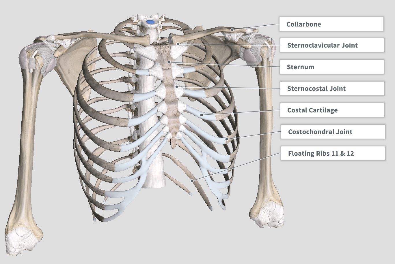
The collarbone (clavicle) links our shoulder blade to our sternum, and the ribs connect to the sternum through sterno-clavicular joints- except for ribs 11 & 12 which are 'floating ribs'. Complete Anatomy Version 2.3.7 (4120), 2017 (1)
There are several layers of muscle in the thoracic spine that allow it to function. Deeper muscle layers help to control movement of adjacent vertebral segments (Inter-transversari, rotatores, multifidus) and to suspend and connect the ribs for functions in breathing and stability (levator costorum, subcostales, intercostals, transversus thoracis and the diaphragm). As well as allowing us to breath, contraction of the diaphgram can generate intra-thoracic pressure and give the thoracic spine stability.
Deep thoracic muscles; back view (left) and front view (right). Complete Anatomy Version 2.3.7 (4120), 2017 (1)
The overlying muscles in the thoracic region span across multiple ribs and vertebrae, helping to generate force and movement. These include the erector spinae muscle group and posterior serratus muscles on the back, and the abdominal wall muscles on the front. These are powerful muscles involved in large movements. The abdominal muscles can also add stiffness and stability to the thoracic spine by anchoring the ribs and generating intra-abdominal pressure (like an internal balloon of air).


Large overlying muscles; back view (left) and front view (right). Complete Anatomy Version 2.3.7 (4120), 2017 (1)
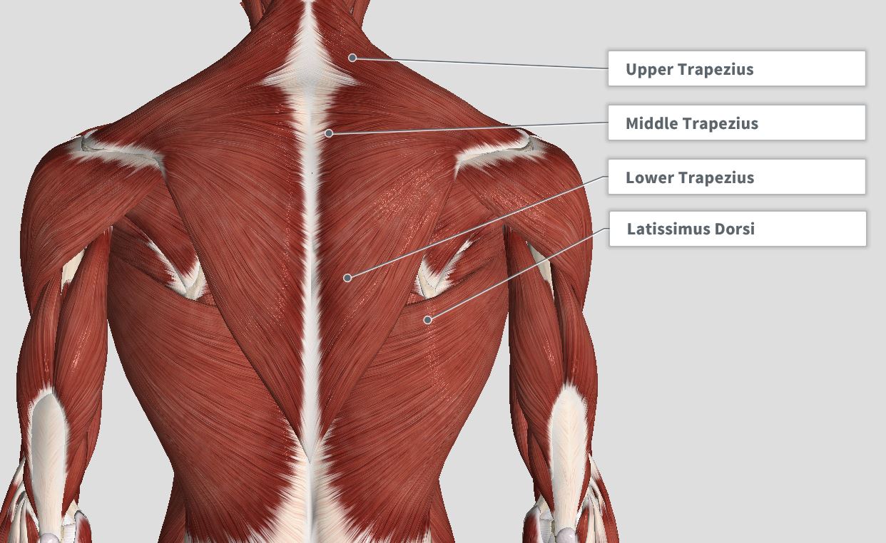
There are also the muscles that connect the shoulder blades (scapulae) and arms to the underlying rib cage and spine. These muscles function to provide a stable base and movement for the upper limbs - as well as postural support and stability in the thoracic region where they help to hold the spine upright and the shoulder blades retracted. In the back, these muscles include the rhomboids, levator scapulae and latissimus dorsi, and the overlying trapezius muscles.

Shoulder blade muscles showing deeper muscles (left) and overlying muscles (right). Complete Anatomy Version 2.3.7, 4120 (2017) (1)
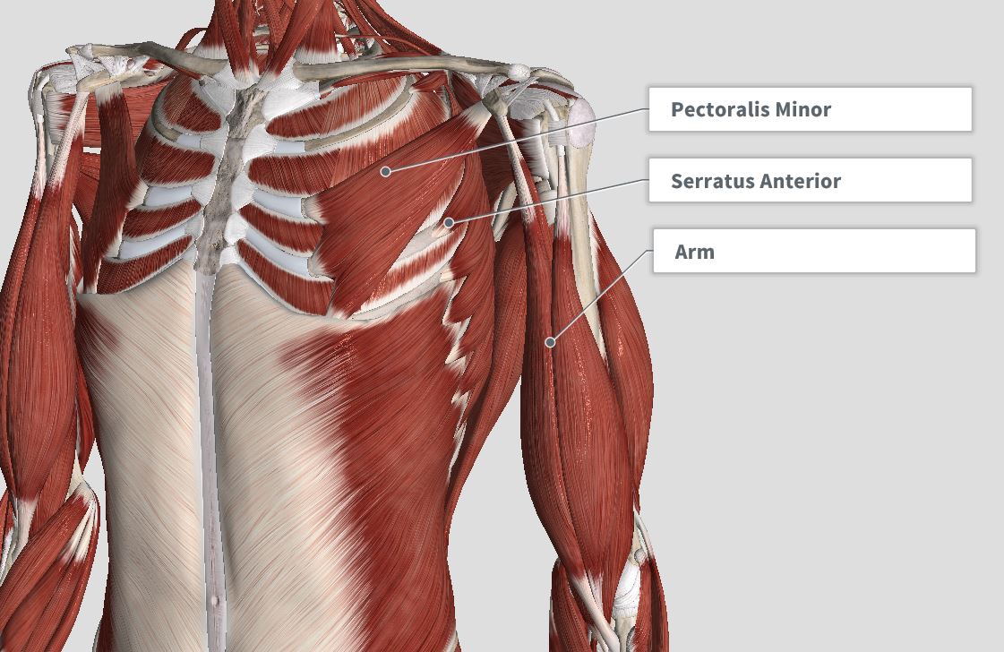
On the front there are the serratus anterior, pectoralis minor muscles and the overlying pectoralis major muscle. In the thoracic region, proper strength and balance between the anterior (front) and posterior (back) shoulder blade muscles is important for proper posture and movement. A common problem is dominance or tension in the front muscles, (especially the pectoralis muscles), with comparative weakness in the posterior muscles that support the spine and retract the shoulder blades (rhomboids, erector spinae). (For more detail on the anatomy, function and problems of the shoulder girdle, see 'Shoulder Rehabilitation' section).

Anterior chest muscles connecting to the shoulder blades (left) and large overlying pectoralis major muscle (right). Complete Anatomy Version 2.3.7 (4120), 2017) (1)
As well as the muscular tissue surrounding the thoracic spine and shoulder blades, there are also layers of connective tissue that acts like a sort of glue, helping to bind overlying muscle layers together and connect muscle to bone. This tissue is known as fascia. Fascia adds further stability to the thoracic spine and helps to transmit forces for movement. At the end of the middle back this fascia becomes thickened near the surface of the spine , called the 'Thoraco-lumbar fascia'. This structure adds further strength and control in this region of the spine.

Fascia refers to connective tissue that binds together successive layers of bone and muscles. It helps to add further strength and control to the middle back.
Complete Anatomy Version 2.3.7 (4120), 2017 (1)
By viewing the anatomy of the thoracic spine you can appreciate that it is a complex structure that must be able to perform a variety of functional roles. As well as providing mobility to allow us to flex, bend and rotate our body, it allows us to breath, provides a stable base of support for movement of our upper limbs and generates movement of our trunk. Often all these tasks are occurring simultaneously - during day to day activities, recreation and sport - with the dynamic interplay between bones, joints and connective tissues allowing this to occur. It is a fine example of the relation between form and function in the human body.
1. 3D4Medical Ltd. (2017).Complete Anatomy (Version 2.3.7 (4120)) [Windows Application Software]. Retrieved from http://3d4medical.com
Where Is the Sternum in Relation to the Shoulder?
Source: https://www.thegapphysio.com.au/thoracic-spine
0 Response to "Where Is the Sternum in Relation to the Shoulder?"
Post a Comment